


Home > Discipline introduction > Equipment
Siemens 1.5T Superconductive MRI
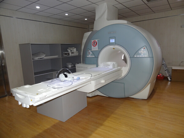
The system has a high-performance gradient system to ensure high signal-noise ratio of the image and high imaging speed. Its multi-system multi-organ characteristic clinical application components are powerful to ensure clear imaging and elegant inspection environment. Its ultra-quiet design reduces noise level by 30dB. Besides, almost all parts can be scanned in the "feet-first" inspection mode, relieving the claustrophobic feelings of the patients and ensuring the most friendly inspection experience throughout the process. So far as the most advanced 1.5T MRI in the industry, it represents the future direction of MRI development, and creates a new field of clinical application for general systemic disease diagnosis.
Ongoing inspection items:
1. Cerebral diseases: MRI is obviously better than CT in displaying and diagnosing cerebral diseases with high image contrast and resolution, direct display from sagittal, axial and coronal planes, without no bone artifacts. And functional MRI: DWI, MRA, MRS, SWI
2. Spinal and spinal cord diseases: MRI is an important method to inspect the spinal and spinal cord diseases. Especially to inspect intraspinal lesions and spinal cord lesions, MRI should be the preferred inspection method.
3. Head, neck and facial diseases: MR can achieve multi-directional scanning with no interference from bone artifacts and high soft tissue resolution, so it is better than CT in displaying lesions in eye, sinus, inner ear, nasopharynx, throat and neck.
4. Abdomen and pelvic cavity: MRI can show iron and other paramagnets, distinguish lipid from aqueous tissues and achieve single tissue imaging, such as magnetic resonanced cholangio-pancreatography (MRCP), magnetic resonance urography (MRU) and the like.
5. Lungs and mediastinum: for lung lesions, CT should be preferred, but for hilar lesions, pleural lesions, lesions near mediastinum and chest wall, MRI can be used. MRI is the preferred examination method to diagnose mediastinal tumors and tumor-like lesions.
6. Bone joint and soft tissue lesions: MRI is the preferred examination method for joint and soft tissue lesions and certain bone lesions, and it is better than CT. Especially in displaying intra-articular structures such as the knee meniscus and cruciate ligament, MRI can be compared with arthroscope. MRI applies to diagnosing soft tissue and bone tumors, inflammation and other diseases.
Unit Level Three
| Financial Category | Code | Test item | Charge unit | Ceiling price (yuan) |
| D | 210200001 | plain MRI scan | ||
| D | 210200001a | plain MRI scan (under 0.5T excluding 0.5T) | Each part | 500 |
| D | 210200001b | plain MRI scan (0.5T-1T) | Each part | 600 |
| D | 210200001c | plain MRI scan (above 1T excluding 1T) | Each part | 750 |
| D | 210200001d | plain MRI scan (3T and above) | Each part | 850 |
| D | 210200002 | Contrast enhancement MRI | ||
| D | 210200002a | Contrast enhancement MRI (under 0.5T excluding 0.5T) | Each part | 550 |
| D | 210200002b | Contrast enhancement MRI (0.5T-1T) | Each part | 650 |
| D | 210200002c | Contrast enhancement MRI (above 1T excluding 1T) | Each part | 800 |
| D | 210200002d | Contrast enhancement MRI (3T and above) | Each part | 950 |
| D | 210200003 | Functional MRI | Time | 550 |
| D | 210200004 | Magnetic resonance cardiac function tests | Time | 550 |
| D | 210200005 | Magnetic resonance angiography (MRA) | Each part | 550 |
| D | 210200006 | Magnetic resonance hydrography (MRCP, MRM, MRU) | Each part | 550 |
| D | 210200007 | Magnetic resonance spectroscopy (MRS) | Each part | 550 |
| D | 210200008 | Magnetic resonance spectroscopy imaging (MRSI) | Time | 550 |